Exploration of Leptospira: A Comprehensive Guide from Understanding to Prevention & Treatment
Leptospirosis is a globally widespread zoonosis. Its causative pathogen poses substantial health hazards to companion animals and may turn fatal under severe conditions. Being contagious, the disease endangers not only pets but also humans, creating potential risks to public health. Hence, knowledge about leptospirosis is critical to safeguard pets and family members.
Pathogen Introduction
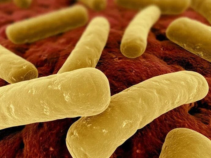
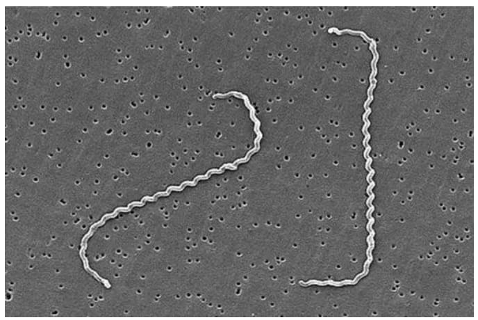
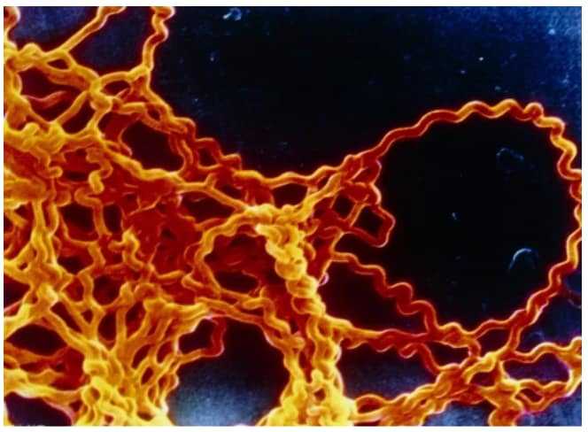
Leptospirosis is triggered by Leptospira, abbreviated as leptospires. Classified as spirochetes, leptospires feature a thin cylindrical body with fine, regular spirals; one or both terminal ends bend into hooks, rendering the organism C-shaped, S-shaped or question-mark-shaped morphologically.
From exterior to interior, the cellular structure consists of outer membrane, cell wall and cytoplasm enclosed by cytoplasmic membrane. Two axial filaments lie between the outer membrane and peptidoglycan layer, each extending from a terminal end toward the cell center.
Leptospires resist conventional Gram staining; Fontana silver impregnation staining is routinely applied to stain the organisms brownish. Typical dimension: roughly 0.1 μm in diameter and 6–20 μm in length. They form right-handed spirals with an amplitude of 0.1–0.15 μm and a wavelength of approximately 0.5 μm.
Leptospira has specific environmental requirements for survival. It thrives in moist soil and water at a pH range of 7.0–7.5 and can survive from several weeks to months during summer, which greatly facilitates its spread. Nevertheless, leptospires are vulnerable to harsh external conditions and die within minutes under dry surroundings. They are susceptible to common disinfectants including 0.5% lysol, 0.1% carbolic acid and 1% bleaching powder, as well as antibiotics such as penicillin and doxycycline.
Epidemiology
Leptospirosis is distributed globally and prevails predominantly in tropical and subtropical zones. In China, the disease has been documented across 28 provinces, municipalities and autonomous regions, with severe epidemics concentrated in rice-growing areas of Central South, Southwest and East China. Rodents and swine constitute the primary infection sources of Leptospira. After contracting leptospirosis, infected rodents and pigs seldom develop obvious adverse impacts on survival, yet their kidneys carry pathogens persistently and continuously shed bacteria via urine, making them the major reservoir hosts and infection sources. Pathogen-containing urine and excreta contaminate the surrounding environment, creating hidden risks for disease transmission.
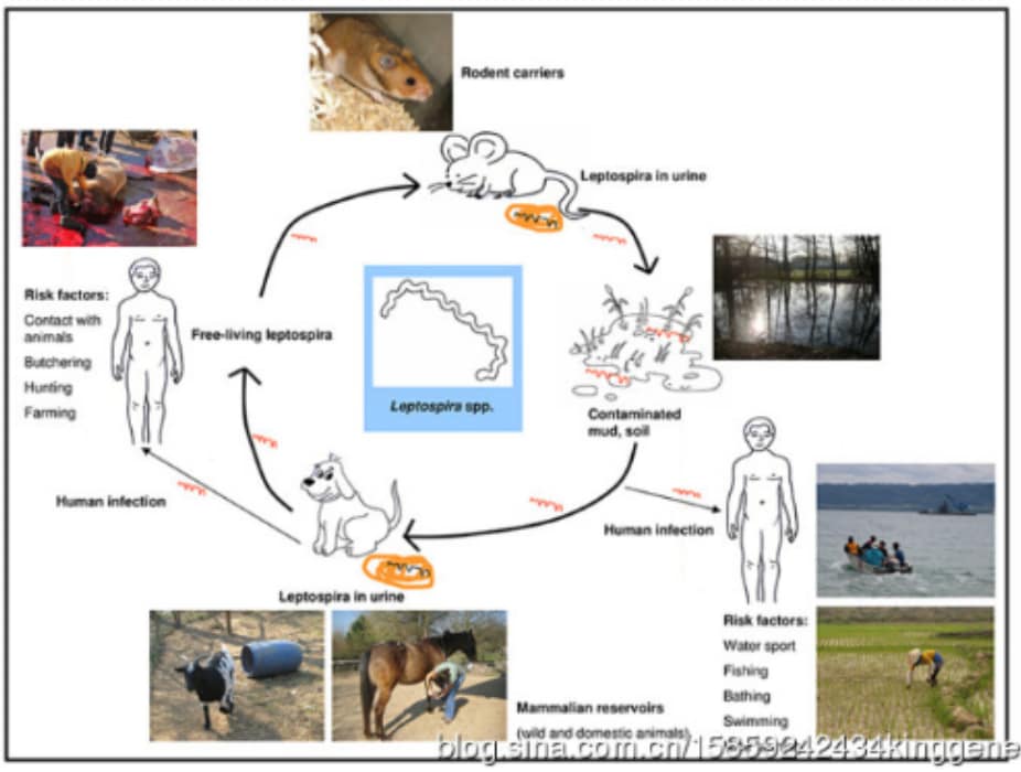
Transmission Routes
Contact transmission: Pets get infected with Leptospira via contact with urine, blood or tissues of infected animals, as well as contaminated soil and water.
Oral transmission: Ingestion of contaminated water or food can also lead to infection in pets.
Transplacental transmission: Pregnant dogs and cats infected with Leptospira may pass the pathogen to fetuses through the placenta.
Nearly all pet species are susceptible to leptospirosis, while high-risk groups include:
- Outdoor pets: Dogs roaming in bushes and woodlands have higher exposure to contaminated surroundings.
- Unvaccinated pets lacking specific immunity against Leptospira.
- Pets residing in rural or suburban areas with abundant pathogen reservoirs.
Clinical Signs
After pets are infected with Leptospira, early manifestations include fever, lethargy, conjunctival congestion and calf muscle tenderness. In the intermediate stage, meningeal signs occur: severe headache, recurrent vomiting, neck stiffness and meningeal irritation. Most pets recover in the late phase, yet some develop permanent sequelae of neurological damage. Humans infected with leptospirosis present similar clinical signs and may develop severe complications such as jaundice, haemorrhage, hepatic and renal failure.
Early-stage signs:
Pets initially display flu-like symptoms including fever, inappetence, depression, vomiting and diarrhoea. These symptoms are commonly misjudged by pet owners as ordinary cold or gastrointestinal upset and easily overlooked.
Intermediate-stage signs:
As disease progresses, severe manifestations emerge: jaundice (yellow discoloration of skin and ocular mucosa), renal failure, liver failure, dyspnoea, myalgia and convulsions, indicating severe organic injury induced by Leptospira.
Late-stage signs and sequelae:
Delayed treatment may trigger disease deterioration and even death. Even after clinical recovery, affected pets are prone to long-term sequelae: renal insufficiency, impaired eyesight and arthralgia.
Diagnostic Methods
The diagnosis of leptospirosis relies on comprehensive evaluation combining epidemiological history, clinical manifestations and laboratory test results.

Serological Tests
Microscopic Agglutination Test (MAT): One of the most widely used assays for leptospirosis. It detects serum antibody levels in pets to confirm infection. Antibodies develop days after exposure, limiting its reliability for early-stage diagnosis. ELISA: Enzyme-linked immunosorbent assay enables rapid antibody screening and aids early diagnosis, yet accuracy is affected by animal age, physical condition and vaccination history. Indirect Immunofluorescence Assay: High sensitivity and specificity for early diagnosis. Result is positive when antibody titer>1:100, or convalescent-phase titer rises ≥4-fold compared with acute-phase serum. Indirect agglutination assays: Diagnostic criteria: single serum indirect hemagglutination titer>1:160, indirect carbon agglutination>1:640, latex agglutination>1:2; or ≥4-fold titer increase in convalescent sample versus acute sample.
Pathogenic Examination
Blood culture: Direct isolation of Leptospira from whole blood; lengthy incubation, rigorous lab requirements and low positive rate restrict routine application.
Molecular Testing(PCR): Rapid and precise detection of leptospiral nucleic acid, ideal for early infection confirmation. Real-time fluorescent quantitative PCR is currently among the most convenient and sensitive detection methods.
Treatment Protocol
The core treatment principle for leptospirosis follows the rule of “three earlies plus on-site therapy”: early detection, early diagnosis, early treatment and local on-site treatment.
Antibiotic Therapy
Penicillin is the first-choice drug to kill leptospira and relieve clinical signs in pets. Standard dosage: 40,000–80,000 IU per kilogram of body weight, intramuscular injection twice daily. It can be combined with streptomycin at 10–15 mg/kg body weight via intramuscular injection, twice per day. Caution: streptomycin shall be avoided or dosage reduced for patients with renal impairment.
For pets allergic to penicillin, gentamicin or tetracycline are alternative options. Ampicillin, amoxicillin and enrofloxacin also deliver satisfactory therapeutic effects; respective dosages are listed below.
| Drug | Recommended Dosage |
| Ampicillin | Canine: 20–30 mg/kg, oral or intramuscular injection, 2–3 times daily. |
| Amoxicillin and Clavulanate Potassium Suspension (Synulox) | Dogs & Cats: 0.1 mL/kg, intramuscular or subcutaneous injection, once daily. |
| Enrofloxacin (Baytril) | Dog: 2.5–5 mg/kg, oral / subcutaneous injection / intravenous infusion, twice daily.Cat: 1–2.5 mg/kg, oral administration, twice daily. |
Symptomatic Treatment
Symptomatic treatment targets specific clinical signs to relieve discomfort of sick pets.
- Fluid, glucose and alkali supplementation: To alleviate gastrointestinal symptoms, administer mixed intravenous infusion of 5% glucose saline (200–500 mL) plus 5% sodium bicarbonate injection (10–40 mL).
- Hemostasis: For bleeding signs, intramuscular injection of vitamin K₁ (2 mg/kg BW) or vitamin K₃ (1 mg/kg BW), twice daily.
- Antiemetic therapy: Metoclopramide at 2 mg/kg BW via intramuscular injection, or oral domperidone 2 mg/kg BW, twice per day.
- Mucocutaneous ulcer care: Iodine glycerin topical application for skin and mucous membrane ulcers.
Precautions
- Early intervention: Timely diagnosis and early medication are critical for leptospirosis management. Take pets for veterinary examination promptly once suspicious signs emerge.
- Medication compliance: Owners must follow veterinary prescription strictly; never adjust dosage or suspend treatment arbitrarily.
- Diet & rest management: Ensure sufficient rest and nutritional supply during treatment. Feed light, easily digestible feed; avoid greasy and irritant food.
Preventive Measures
Leptospirosis prevention shall be implemented from the following aspects:
1. Block transmission routes & control infection sources
- Carry out rodent prevention and extermination, properly manage livestock excreta to avoid contamination of water sources and feeds.
- Regular parasitic deworming for pets: Ectoparasites such as fleas and ticks can act as leptospira transmitters; routine deworming helps cut parasitic-borne infection.
- Restrict pets’ access to wild animals especially rodents and pigs that likely carry pathogens.
2. Environmental sanitation management
- Regularly clean pet living areas; disinfect premises with chlorine-containing disinfectants or ultraviolet irradiation to inhibit pathogen proliferation.
- Prevent pets from drinking raw unboiled water; supply clean drinking water exclusively.
- Bathe pets periodically with mild pet-specific shampoo; refrain from irritant detergents to keep body hygiene.
3. Additional protection strategies
- Vaccination: Leptospira vaccination is the most effective preventive method. Follow veterinary schedules for regular inoculation to boost specific immunity.
- Improve physical fitness via balanced diet and proper exercise to strengthen pets’ general disease resistance.
Regular Physical Examination
Periodic PCR testing for Leptospira enables early and accurate detection of pet infection status, facilitating preventive care for healthy animals and prompt treatment for infected individuals.
Leptospirosis is a severe zoonotic disease. Nevertheless, scientific treatment and effective preventive measures can drastically reduce infection risks. All pet owners are advised to attach importance to their pets’ health, arrange regular physical check-ups and scheduled vaccinations at veterinary clinics, so furry companions can grow up healthy and happy. Meanwhile, proper personal protection is essential to prevent infection of owners and family members from pets.