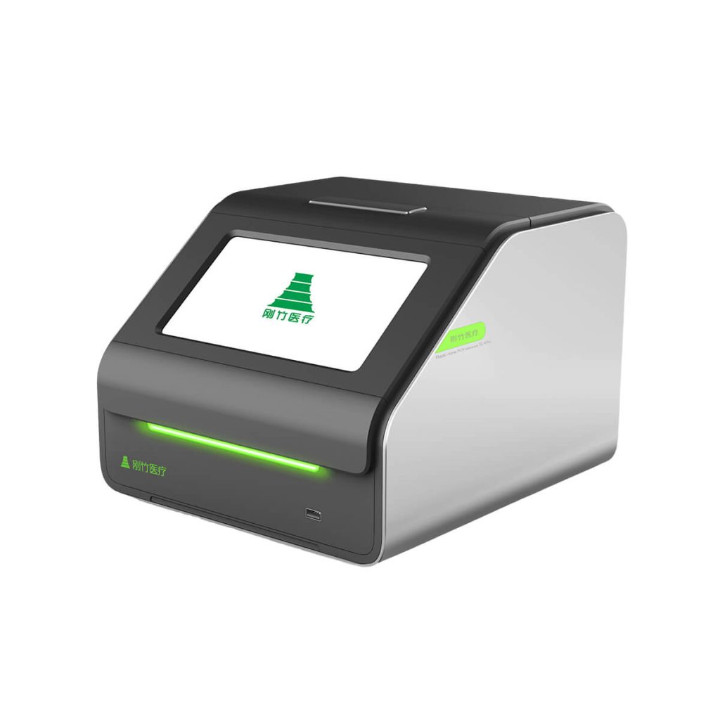
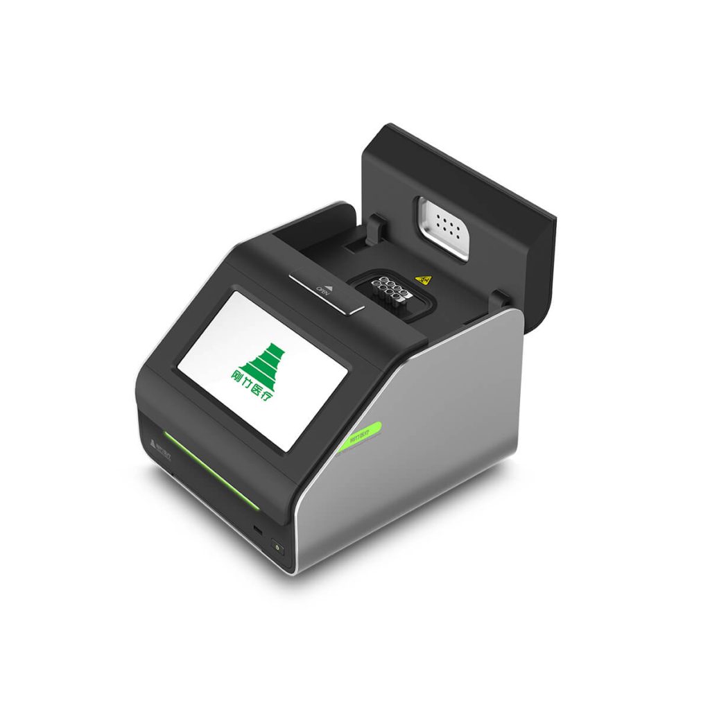
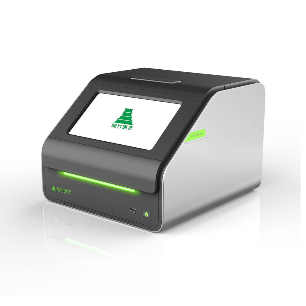
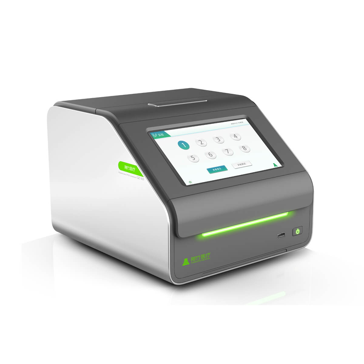
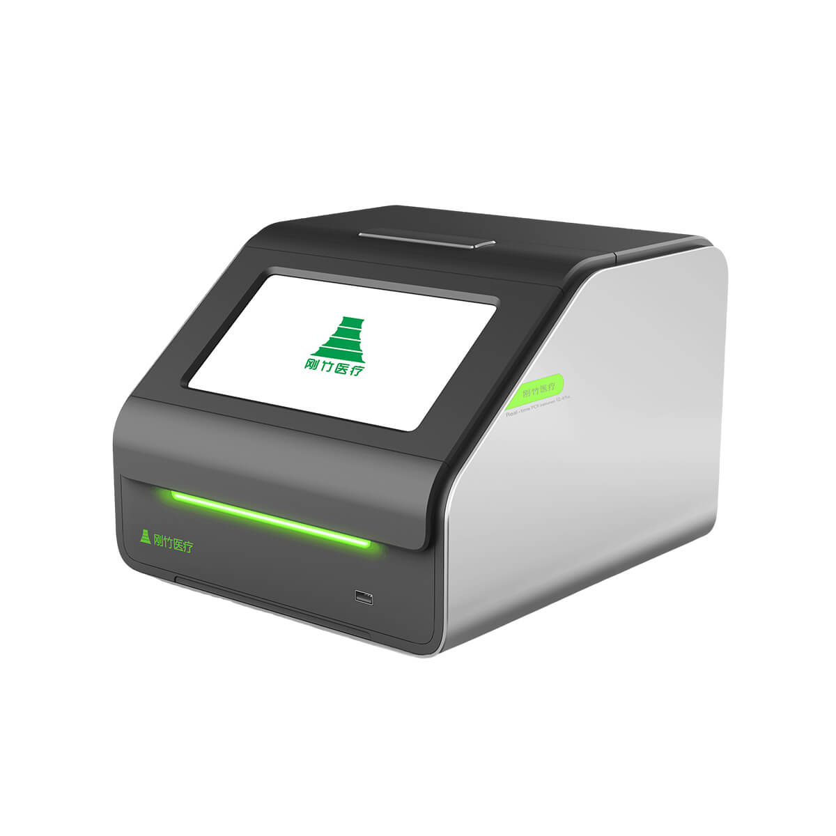
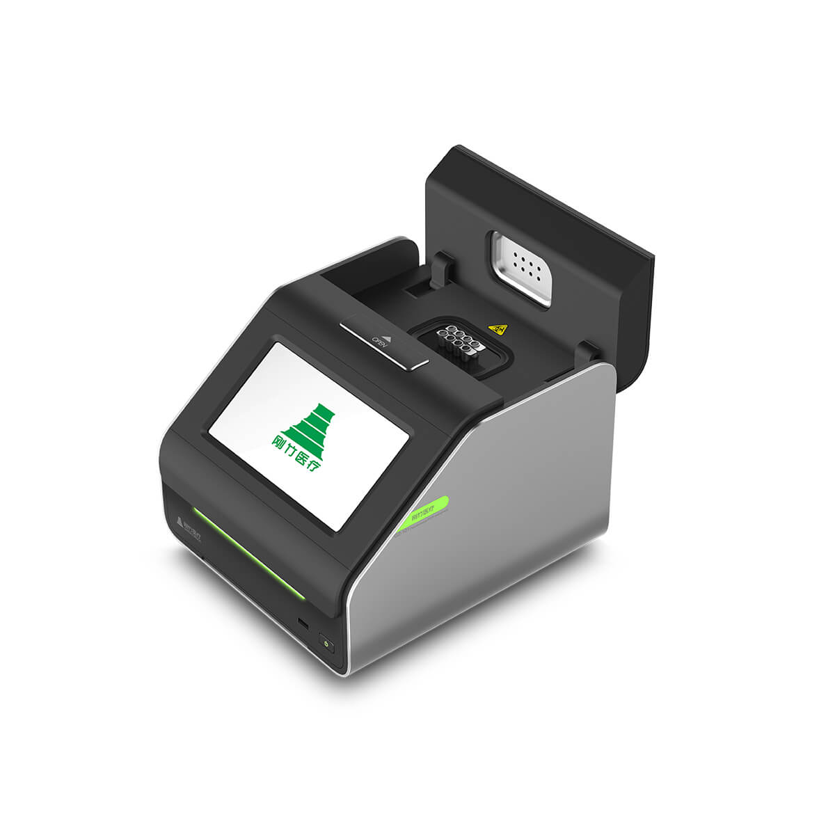
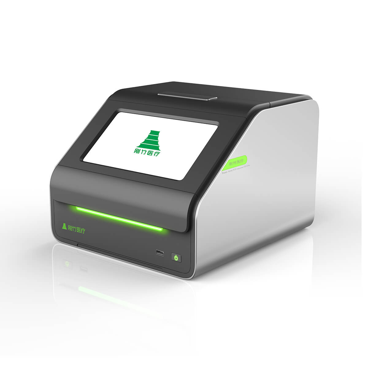
GZ-8Plus便携式实时荧光PCR仪

为宠物医院提供宠物传染疾病诊断解决方案是疾病确诊的首选产品

  • 同时可测8个样本16各项目
  • 机身尺寸小
  • 精准的温控系统
  • 支持WiFi数据传输
  • 云端数据管理系统
  • 一键生成检测报告
Basic ParametersSize268*236*188 mmPower frequency50Hz-60Hz
VoltageAC 110V/220VCommunication InterfaceWiFi, USB
Weight4.5 KgSample size8 holes*0.2ml
Use EnvironmentTemperature10°℃-35℃Atmospheric pressurestandard atmospheric pressure
Humidity10%-95%RH, no condensationAltitudeBelow 2000 meters
Fluorescence Detection System PerformanceLight sourceHigh brightness LEDDetectorPD
Sample Linear Range10-1E10 copySample LinearityR≥0.99
Thermal ParametersTemperature accuracy±0.25℃Temperature control accuracy±0.1°℃
Temperature uniformity±0.25°℃Heating and cooling rate6°C/S
Optical ParametersExcitation wavelength of the first channel495nm±10nmExcitation wavelength of the second channel535nm±10nm
Detection wavelength of the first channel520nm±10nmDetection wavelength of the second channel570nm±10nm

Step 1: Put 20ul of extracted nucleic acid liquid into the test reagent tube.

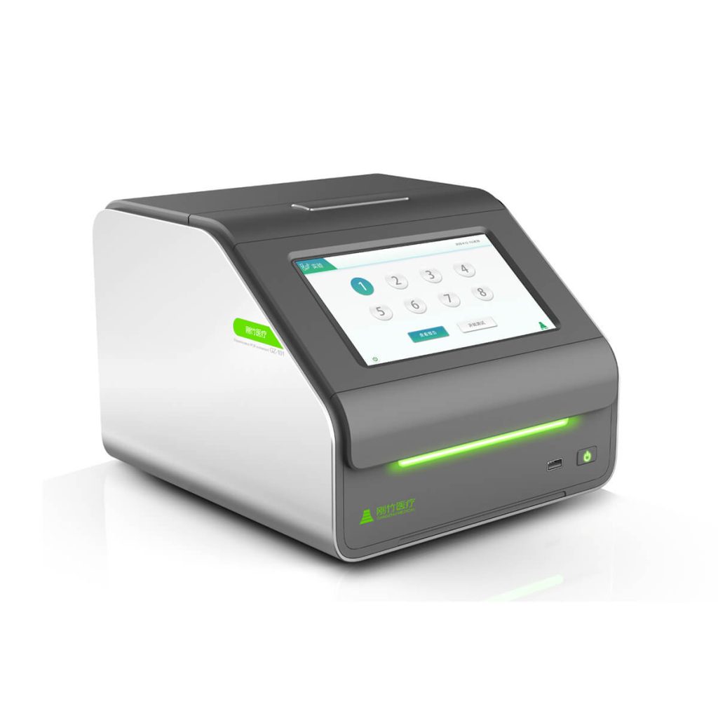
Step 2: turn on the GZ-8plus instrument, connect wifi, then the screen will show 1-8 wells numbers like below:

Step 3: Choose a well to put the reagent tube in, and click the number of matching wells. Then type in the sample & other information below the interface.

Step 4: Click Run to start the test or put other reagent with samples in rest wells and run test.

Canine & Feline Pathogens

Concurrent Illnesses in Humans and Pets

Exotic Animal Pathogens

Animal health Genetic test: Tumor, Kidney disease, Blood Type(developing)

Consumer-Grade Genetic Testing for Pets(developing)
Clean and maintain the PCR machine: Use 75% alcohol or wet wipes to first clean the external surfaces, removing dust and stains. Then, dry the surfaces with a dry tissue.

(Note: When cleaning and wiping the hot cover and reaction hole, be sure not to drip liquid into the hole to avoid damaging the internal components.)

PCR machine: Observe if the running indicator lights are normal, check the running interface countdown, temperature display, Lid, and amplification curve for normal change.

During normal operation, the amplification curve should start to appear around 40 minutes into the countdown. If not, there may be an issue with the operation. Please contact customer support promptly.

The Ct value represents the number of amplification cycles during the PCR process at which the fluorescence signal of the amplification product reaches the set fluorescence signal threshold.

The Ct value is inversely proportional to the initial template concentration in the PCR reaction. The larger the Ct value, the lower the initial template concentration. In other words, a higher Ct value indicates a lower viral content in the sample. For different testing projects, the clinical significance of Ct values may vary. For instance, for certain viruses like the influenza virus, a Ct value above 26 indicates a very low viral content, and many cases may present as asymptomatic carriers.

A negative nucleic acid test means that no nucleic acid of the relevant pathogen was detected in this experiment; a positive test means that the nucleic acid of the relevant pathogen was detected in the sample.

When a nucleic acid test is positive, if the pet shows no symptoms, it is considered asymptomatic carrier.

When the nucleic acid test is positive and the pet exhibits symptoms, the viral infection may be related to the symptoms. However, a comprehensive assessment of the pathogenic factors should be made by considering the Ct value (viral load level) and other test results.

(Note: A single positive result in nucleic acid testing cannot be used as the sole basis for direct diagnosis, to avoid misdiagnosis.)

The iconic feature of fluorescence quantitative PCR detection is the PCR amplification curve. Whether the result is valid can be judged based on whether the linear change of the amplification curve conforms to the standard amplification linear change trend.

(Note: The standard amplification exhibits an S-shaped smooth curve trend, with a certain increase in fluorescence signal. The original curve is generally consistent with the amplification curve’s basic changes.)

If a pet shows clear symptoms, and all results from relevant testing projects are negative, considerations and analysis can be made from the following aspects:

  1. Investigate whether there is a possibility of operational errors during the testing process, such as accurately measuring the sample volume or ensuring the proper use of reagents.
  2. Examine whether the sampling was done correctly, and additionally, check if excessive samples were diluted before nucleic acid extraction.
  3. Check if the reagents used are normal, if there are any missing reagents, and whether the lyophilized powder reagents are in an intact white lump-like state.
  4. Assess whether the temperature during the nucleic acid extraction process, including the temperature of the extraction reagents and the laboratory environment, was within the recommended range of room temperature (20-35℃).
  5. Evaluate whether the instrument operated normally during the process.

If all the above issues are ruled out, it may be that the pathogen is indeed not present in the sample or its concentration is below the detection limit. It’s possible that the detected pathogen is not the main cause of the observed symptoms.

The situation where the two test results are inconsistent (both are the results of normal testing operations) can generally be divided into two situations:

The first situation is different results from samples collected at different times: Because the sampling time and techniques may vary, and after undergoing two rounds of nucleic acid extraction and PCR amplification detection processes, especially for weak positive cases where the virus content is low, there may be unavoidable less-than-ideal repeatability, leading to inconsistent results in two consecutive tests.

The second situation is different results from two consecutive tests on the same sample: Excluding instrument malfunctions, reagent failures, and human operational errors, it is generally possible that these are borderline cases. The differences in nucleic acid extraction efficiency and reaction amplification efficiency during each test may lead to inconsistent results in the final two tests.

As a primary principle, the pipette and its tips should not be mixed with others and should be used independently.

Maintain proper usage of the pipette to avoid liquid backflow. It is recommended to clean the pipette with 75% alcohol before and after each experiment.

Keep the pipette tips clean inside the box for proper use. It is recommended to replace with new sterile tips regularly (monthly), either by purchasing new boxed tips or by using sterile gloves to insert new tips into a clean tip box. Additionally, after each use, promptly cover the box to prevent contamination.

Wearing gloves or sealing waste is crucial for preventing nucleic acid contamination. It is essential to pay attention and avoid neglecting these measures.

Notice: Wear disposable, powder-free latex/nitrile gloves when conducting experiments! Sampling gloves cannot be used directly for experimental operations; they must be replaced with new gloves. When opening the reagent tube cap each time, be sure to avoid touching the inner side of the cap. If accidentally touched, promptly replace with new gloves or clean the contaminated area of the gloves with 75% alcohol.

All discarded pipette tips, waste liquids, residual samples, PCR amplification products, etc., must be sealed in self-sealing bags and cannot be left open.