
Rickettsia rickettsii, another tick-borne human and pet disease
Canine and feline rickettsial diseases are a group of diseases caused by microbial infections of the genus Rickettsia, which pose a significant threat to the health of pets and, in some types, a potential risk of human-pet co-infection. In recent years, with the popularization of pet ownership and the development of veterinary medicine, Rickettsia infections have been increasingly studied. The purpose of this article is to provide a comprehensive analysis of the impact of rickettsia in pets, from pathogen overview to preventive measures, and to provide a detailed reference guide for veterinarians and pet parents.

Pathogen Overview
Rickettsia rickettsii belongs to the family Rickettsiaceae, which can be categorized into spotted fever group Rickettsia felis and ornithosis group Rickettsia rickettsii based on their biological characteristics and pathogenicity. The spotted fever group of rickettsiae includes a variety of pathogens capable of causing infection in pets, such as feline rickettsia (Rickettsia felis) and canine rickettsia (Rickettsia rickettsii). Rickettsia felis is mainly transmitted by fleas, and infection can cause fever, rash and other symptoms in cats, and in severe cases, can cause multiple organ damage. Canine rickettsia rickettsii is transmitted by ticks, and infection of canines can cause fever, arthritis, neurological symptoms, etc., and can lead to death in severe cases. Rickettsia ornithosis mainly infects birds and has a relatively small impact on pets.
Rickettsiae are microorganisms that are intermediate between bacteria and viruses and have strictly intracellular parasitic properties. They are mainly transmitted by arthropods such as fleas, lice and ticks and cause a wide range of infectious diseases. Rickettsiae are usually between 0.2 and 0.5 micrometers in size and have a variety of morphologies, including spherical and rod-shaped. Its cell wall structure is similar to that of bacteria. It has a cell wall, which is made up of lipopolysaccharides and proteins, and contains both RNA and DNA inside the cell, which are categorized as Gram-negative bacteria. It is sensitive to a variety of antibiotics, such as tetracyclines and chloramphenicol.
Epidemiology
Rickettsial diseases are characterized by marked seasonal, regional and population susceptibility. The incidence of rickettsial disease is higher during warm and humid seasons due to high arthropod activity. In addition, epidemiologic investigations have shown that rickettsial diseases occur globally and are particularly common in tropical and subtropical regions. Factors such as a pet’s range of motion, exposure to the environment, and frequency of contact with wildlife can also influence the prevalence of rickettsial disease.
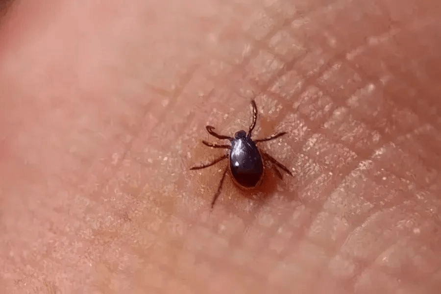
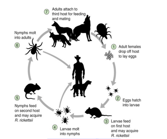
The main mode of transmission of rickettsial infection in pets is arthropod bites. Like ticks, fleas, lice, and mites, they feed on the blood of animals infected with Rickettsia, the pathogen multiplies in their bodies, and then spreads the pathogen when they bite a healthy pet. For example, canine Ehrlichia is often transmitted through brown dog ticks, and feline Rickettsia is mostly carried by cat fleas. The risk of infection varies among pets of different breeds, ages, geographic regions, and seasons. Purebred dogs such as German shepherds and Rottweilers are at higher risk of canine ehrlichia compared to mutts; young and old pets with weakened immunity are more likely to be infected than adult pets.
In addition to this, Rickettsia can be spread through contact with infected pets’ feces, urine and other secretions, or through contact with contaminated environments and objects. Some rickettsial infections are zoonotic and pose a threat to human health as well. Therefore, the control and elimination of these vectors is an important measure in the prevention of rickettsial diseases.
Infection Cycle & Pathogenesis
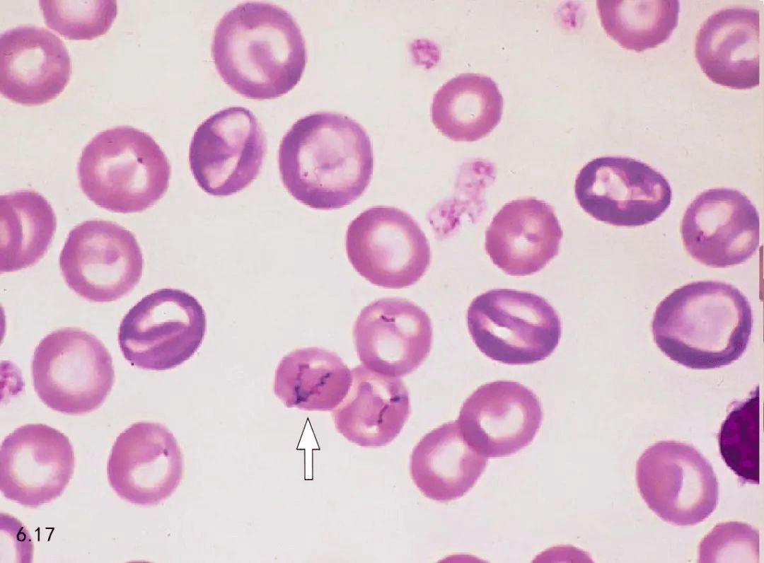
The infection cycle of Rickettsia rickettsii consists of several stages. First, when a vector such as a flea or tick bites an infected host, Rickettsia enters the vector and multiplies in its digestive tract. Subsequently, when the vector bites a healthy pet again, the rickettsiae enter the pet through saliva or feces. Upon entering the pet’s body, rickettsiae invade the endothelial cells of blood vessels, multiply within the cells and release toxins, leading to vascular inflammation and tissue damage. After infection, pets develop clinical signs such as fever, rash, and arthritis. As the disease progresses, Rickettsia can further spread to organs throughout the body, causing multi-system pathology. The length of the infection cycle varies depending on the type of rickettsiae and host immunity, usually between 1-3 weeks.
Once rickettsia enters the body, it spreads through the lymphatics or directly into the bloodstream to small capillaries, where it primarily infects endothelial cells, and damage to endothelial cells leads to vasculitis and increased microvascular permeability. Mechanisms of cellular injury include oxidative damage through the production of reactive oxygen species, cellular necrosis, and induction of endothelial cell apoptosis by CD8+ T cells. Endothelial cell death, the presence of inflammatory cytokines (e.g., IL1-β, IFN-γ, and TNF-α) as well as the induction of COX-2 and subsequent prostaglandins combine to effect an increase in vascular permeability thereby causing tissue edema.
Clinical symptom
The clinical signs of rickettsial infection in dogs are varied and complex, and multiple clinical signs will also lead to anemia, including the following:
Fever: Most infected dogs will show signs of fever, which can rise to 39.5-41°C. Biphasic fever, i.e., the body temperature first rises, then falls, then rises again for an indeterminate period of time.
Respiratory symptoms: purulent mucus from eyes and nose, sneezing, coughing, and in severe cases, pneumonia, mainly abdominal breathing. This is due to bronchopneumonia and other respiratory diseases caused by rickettsiae.
Digestive tract symptoms: decreased appetite, vomiting, drainage-like or mucus stools, and in severe cases, bloody stools, extreme dehydration and lethargy in sick dogs. These symptoms are due to the attack of rickettsiae on the digestive tract, resulting in gastrointestinal dysfunction.
Neurological symptoms: convulsions and spasms, which may start at the corners of the mouth or the limbs and gradually worsen, ending in convulsions of the limbs or the whole body. In severe cases, neurological symptoms such as epilepsy may occur, due to damage to the central nervous system by rickettsiae.
Skin symptoms: skin keratoses, such as keratotic dryness of the nose and keratinization of the pads of the feet to form scleroderma. Skin lesions such as rashes may also occur.
Eye damage: clinically characterized by keratitis, conjunctivitis, whitening of the cornea, and in severe cases, perforation, corneal ulcers, and blindness. This is due to the infection and destruction of ocular tissues by rickettsiae.
When cats are infected with Rickettsiae, their clinical symptoms are different from those of dogs, as follows:
Fever: infected cats generally show fever symptoms and elevated body temperature, but the specific temperature range and duration may vary according to individual differences.
Digestive symptoms: loss of appetite, vomiting, diarrhea, and in severe cases, blood may be present in the feces, which is coffee-colored. This is due to the damage caused by Rickettsia rickettsii to the cat’s digestive tract, triggering diseases such as gastroenteritis.
Respiratory symptoms: Some infected cats will show respiratory symptoms such as coughing and runny nose, but compared to dogs, respiratory symptoms in cats are usually milder.
Eye symptoms: Conjunctivitis is more common, and the eyes will be red, swollen and watery. In severe cases, this can lead to corneal damage.
Skin Symptoms: Rash is an important manifestation of rickettsial infection in cats, with red spots, pimples and other lesions on the skin.
Anemia: Due to anemia, the conjunctiva of the eyes of sick cats may become pale.
Systemic Symptoms: Systemic symptoms such as depression, decreased activity and weight loss are also more pronounced, reflecting the deterioration of the cat’s overall health.
Symptoms of feline rickettsial infections are similar to those of other rickettsial and viral infections and are more difficult to differentiate clinically, thus their incidence may be underestimated due to misdiagnosis.
Diagnostic methods
The diagnosis of rickettsial infection requires a combination of factors. First of all, based on the pet’s clinical symptoms such as fever, dysphoria, anemia, respiratory or gastrointestinal symptoms, can be initially suspected that the infection may be possible, but these symptoms are not unique to rickettsial infections, as canine distemper, microvirus infections and other diseases have a similar manifestation, so can not be based on the symptoms alone to confirm the diagnosis. Laboratory testing is a key step in confirming the diagnosis and includes the following methods:
Serologic testing: Diagnosis of rickettsial infection is made by detecting specific antibodies in the pet’s serum. Commonly used methods include enzyme-linked immunosorbent assay (ELISA) and immunofluorescence. The advantage of serologic testing is that it is relatively easy to perform, but it requires a window of time and antibodies may not be detected in early infections.
Molecular biology testing: Rickettsia rickettsii DNA is detected using polymerase chain reaction (PCR) technology. this method is extremely sensitive and specific, and can detect the presence of the pathogen in the early stages of infection.The real-time fluorescent quantitative PCR (qPCR) technique also allows for quantitative analysis of the pathogen load, which helps to assess the severity of the disease and the efficacy of treatment.
Pathogen isolation: Blood or tissue samples from pets are inoculated into specific cell culture media to observe the growth of Rickettsiae. This method allows for the acquisition of live pathogens, providing material for further biological studies and drug screening. However, because Rickettsia is a strictly intracellular parasitic microorganism, the culture conditions are more demanding and there is a certain biosafety risk, so it is somewhat limited in practical application.
Direct microscopy: direct observation of rickettsiae through microscope. Commonly used methods include Giemsa staining and immunofluorescence staining. The advantage of direct microscopy is that it can visualize the morphology and distribution of the pathogen, but it requires high requirements for operating techniques and equipment, and needs to be differentiated from other similar pathogens for diagnosis.
Imaging examination: imaging examination plays an auxiliary role in the diagnosis of rickettsial disease, mainly used to observe the lesions of the pet’s internal organs and exclude other diseases.
Treatment recommendations
Antibiotics are preferred for the treatment of rickettsial disease, of which tetracycline antibiotics such as doxycycline are commonly used.
Doxycycline: dogs, cats, internal administration of a single dose of 5-10mg per 1kg body weight, once a day, for 3-5 days, can inhibit the protein synthesis of rickettsiae, hindering its growth and reproducti.
Tulathromycin: Canine intravenous drip once the amount of 10 mg per 1 kg of body weight, 2 times/day; oral once the amount of 20 mg per 1 kg of body weight, 3 times/day, but these two drugs are relatively more side effects, may cause gastrointestinal discomfort, such as vomiting, diarrhea, long-term use may also affect the development of teeth, bones, young pets need to be used with caution.
Sulfonamides such as sulfadimethoxine: 60 mg/kg body weight, oral 3 times/day, or sulfadimethoxine sodium injection, 30 mg/kg body weight, intravenous drip, but note that the first time you need to use a shock amount (2-4 times the normal dose), followed by a normal amount of symptoms relieved by halving the amount of sulfonamide crystals during the period of time with the same amount of sodium bicarbonate alkalinization of the urine, to prevent sulfonamide crystallization damage to the kidneys.
In addition to antibiotic treatment, supportive therapy is critical in the management of rickettsial disease.
For pets with significant signs of fever, physical cooling measures, such as using ice packs on areas rich in large blood vessels, such as the neck and groin, or giving warm water baths, can be used to help lower body temperature. At the same time, antipyretic drugs such as nonsteroidal anti-inflammatory drugs (NSAIDs) can also be given according to the specific conditions of the pet, but attention should be paid to the dosage of the drug and the frequency of its use to avoid causing adverse reactions to the pet.
For pets with gastrointestinal symptoms such as vomiting and diarrhea, hydration and electrolytes should be replenished to prevent dehydration and electrolyte disorders. Pets can be given appropriate amounts of saline or glucose saline orally or intravenously, and electrolyte solutions can be added if necessary.
For pets with reduced appetite, nutritious, easily digestible food, such as fluid or semi-liquid food, can be provided, and nutritional supplements can be added appropriately to enhance the pet’s physical fitness and immunity.
For severe cases, such as respiratory distress, heart failure and other serious complications, appropriate symptomatic treatment such as oxygen inhalation and cardiotonic drugs should be given immediately, and the pet’s vital signs should be closely monitored to ensure its stability.
In the process of supportive treatment, it is also necessary to keep the living environment of pets clean, quiet and comfortable, to avoid their external stimulation and interference, which is conducive to the recovery of pets.
After a period of treatment, regular review is required to assess the efficacy of the treatment through routine blood tests, blood biochemistry, and pathogenetic tests, to see if the rickettsiae are cleared, whether the blood cell indexes are restored, and whether there is any improvement in organ function, and to adjust the treatment based on the results until the pet recovers completely. After timely treatment, most canine and feline patients have a favorable prognosis; however, if serious complications occur, such as multiple organ failure, the prognosis is poorer.
Preventive measures
Regular cleaning and disinfection: Keep the living environment of pets clean and hygienic, and thoroughly clean and disinfect the living space, activity areas and related facilities of pets on a regular basis. Physical prevention and control: Adopt physical methods to eliminate arthropods, such as using vacuum cleaners to thoroughly vacuum the pet’s activity area, which can suck away flea eggs, larvae and adult fleas and reduce their numbers; for larger arthropods such as ticks, tools such as tweezers can be used to manually capture and remove them.
Chemical Control: Environmental spraying with insecticides that are effective against arthropods can be used when necessary. Choose insecticides that are relatively safe for pets and humans, such as pyrethroids.
Regular medical checkups: Take your pet for regular medical checkups to detect and deal with health problems in a timely manner.
Vaccination: Vaccination against rickettsial diseases, such as canine rickettsial disease, can be chosen according to the living environment.
Deworming and pest control: Regularly deworm your pet both internally and externally, especially for fleas, lice, ticks and other vectors.
Enhance nutrition and exercise: Provide pets with balanced nutrition and ensure that they consume enough protein, vitamins, minerals and other nutrients to enhance their physical fitness and immunity. At the same time, pets are encouraged to take moderate exercise, such as walking and playing, to promote their physical health and improve their resistance to diseases.
In addition, pet owners should enhance their awareness of self-protection and avoid direct contact with infected pets to reduce the risk of transmission of zoonotic diseases.
Final Thoughts
Rickettsial disease in cats and dogs is a complex and diverse group of infectious diseases in humans and pets, with hidden transmission and potentially serious harm. The occurrence and spread of rickettsial infections can be effectively reduced through scientific diagnosis and treatment methods and timely preventive measures. May every owner’s careful care keep their pets away from rickettsiae and have a happy and carefree life.