Interpretation of Feline Leukemia Virus (FeLV) Test Results
Introduction
In clinical veterinary practice, dealing with diagnostic reports is routine. Often, veterinarians need
to combine multiple tests to make a diagnosis. However, sometimes “conflicting” situations arise
where two or more test results contradict each other, causing doubt and leading to questions about
the accuracy of testing methods. This scenario frequently occurs with Feline Leukemia Virus
(FeLV). How should such situations be resolved?

To address this challenge, we first need to understand FeLV testing methods and various infection
scenarios:
Testing Methods
Antigen: Generally, ELISA or SNAP methods are used to detect p27 protein. This is because p27
protein is produced in quantities exceeding those necessary for assembling new virus particles,
and p27 is abundant not only in the cytoplasm of individual infected cells but also in the blood of
infected cats. Whole blood/serum can be used. If hemolysis occurs, whole blood samples have a
higher false-positive probability. Due to the design of certain reagents with filtration membranes,
results may differ between whole blood and serum samples. Overall positive predictive value is
50%-80%, with a negative predictive value of 100%.
PCR: Standard RNA detection tests for the virus’s genetic material itself. Proviral DNA detection
tests for host cells. FeLV uses reverse transcriptase to transcribe into proviral DNA, which then
integrates into the host’s bone marrow stem cell genome. Gene fragments from blood cells derived
from stem cell replication and differentiation can be detected in the blood.
Infection Classifications
(1) Abortive Infection: (Immunity >> Virulence)
Typically occurs during initial FeLV infection. Viral replication is generally limited to local
oropharyngeal lymphoid tissues. In cats with strong immunity, the immune system can suppress
viral replication through humoral and cellular immunity, preventing viremia. These cats develop
strong immunity against FeLV that can last for several years. Test Results: High levels of
neutralizing antibodies, but antigen, RT-PCR for RNA, and proviral DNA tests are negative.
(2) Regressive Infection (Immunity > Virulence)
After the virus enters the body, it spreads systemically in lymphocytes and monocytes. The
immune system responds, clearing the virus from the blood but not completely from the body.
Some virus may integrate into bone marrow hematopoietic stem cells. This situation represents a
normal immune response, unlike abortive infection. Due to systemic viral replication, infected cats
may show symptoms such as lethargy, fever, and lymph node enlargement. The virus typically
persists in the blood for 1-6 weeks, rarely for several months, before being cleared. The longer the
virus persists, the harder it is to eliminate. After viremia is suppressed, the virus can be reactivated
if the immune system is compromised. Cats in this state can transmit FeLV through blood
transfusion. Test Results: p27 antigen and RNA tests may be positive or negative (depending on
the clearance time window of viremia or reactivation), proviral DNA test is positive.
(3) Progressive Infection (Virulence > Immunity)
If a cat’s resistance is weak or the viral strain is highly virulent, FeLV replicates widely in the cat’s
body, causing persistent viremia. It first affects lymphoid tissues, then bone marrow, mucosal and
glandular epithelial tissues. Infection of mucosa and glands is related to viral shedding, which is
why saliva from infected cats is infectious. The probability of infection from a shedding cat to a
normal cat is 5%, but for kittens in prolonged contact with shedding cats, the infection rate can
reach 30%. Progressive FeLV infection can lead to tumors, hematological diseases,
immunosuppression, immune-mediated diseases, and other syndromes (including reproductive
disorders and neuropathies). In multi-cat households, the mortality rate for cats with progressive
infection is described as approximately 50% within 2 years and 80% within 3 years. Test Results:
As progressive infection takes time to develop (2-6 weeks), p27 may be positive or negative
before the infection is established, and positive after establishment. PCR tests for proviral DNA
and RNA are both positive.
(4) Local Infection
This is a rare form of infection characterized by persistent local viral replication in certain tissues
such as the spleen, lymph nodes, small intestine, or mammary glands. Test Results: Whole blood
RNA test is negative, p27 and proviral DNA may be positive or negative.
| Testing Method | p27 Antigen | RNA Test Proviral | DNA Test |
|---|---|---|---|
| Abortive Infection | (-) | (-) | (-) |
| Regressive Infection | (+)/(-) | (+)/(-) | (+) |
| Progressive Infection | (+) | (+) | (+) |
| Local Infection | / | / | / |
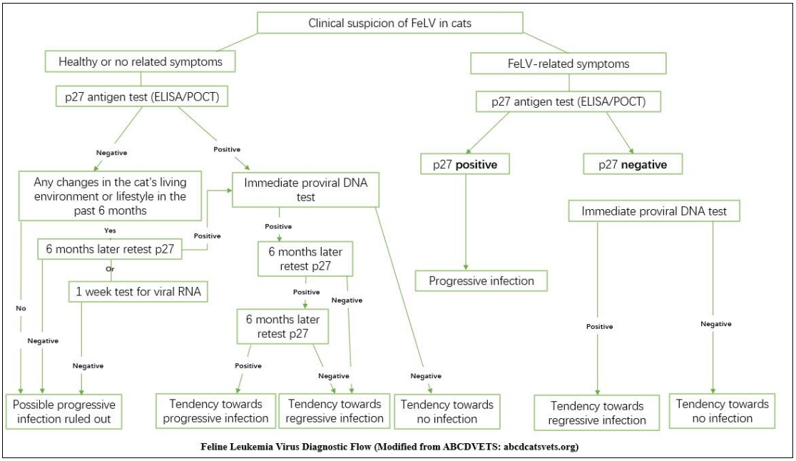
Common Diagnostic Process

Summary
In conclusion, p27 antigen testing can serve as a general screening tool and differentiate between progressive and regressive infections. For asymptomatic cats, negative results are highly reliable. However, for cats with symptoms or suspected FeLV, negative p27 results are somewhat limited and should be interpreted in conjunction with proviral DNA results. Comparatively, proviral DNA testing is an effective means of detecting FeLV infection. A positive result indicates infection (regressive or progressive), while a negative result suggests no infection or abortive infection. For accurate diagnosis and prognosis of feline leukemia virus, beyond correct interpretation of diagnostic methods, long-term follow-up testing is crucial.
References
GREENE’S INFECTTIOUS DISEASES OF THE DOG AND CAT, chapter 30. Hofmann-Lehmann R, Hartmann K. Feline leukaemia virus infection: A practical approach to diagnosis.J Feline Med Surg. 2020 Sep;22(9):831-846. doi: 10.1177/1098612X20941785
